Copyright
©2014 Baishideng Publishing Group Co.
World J Gastroenterol. Feb 14, 2014; 20(6): 1630-1634
Published online Feb 14, 2014. doi: 10.3748/wjg.v20.i6.1630
Published online Feb 14, 2014. doi: 10.3748/wjg.v20.i6.1630
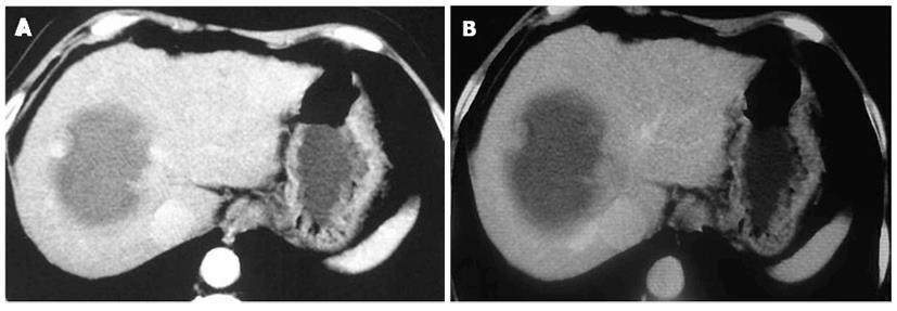
Figure 3 Contrast-enhanced computed tomographic of a primary hepatic carcinosarcoma.
A: Contrast-enhanced computed tomographic revealed peripheral nodular enhancement in the arterial phase with a large internal non-enhancing portion, which correlated with contrast-enhanced ultrasonography findings; B: Peripheral nodular portion of the tumor was isoenhanced in the portal phase.
- Citation: Liu LP, Yu XL, Liang P, Dong BW. Characterization of primary hepatic carcinosarcoma by contrast-enhanced ultrasonography: A case report. World J Gastroenterol 2014; 20(6): 1630-1634
- URL: https://www.wjgnet.com/1007-9327/full/v20/i6/1630.htm
- DOI: https://dx.doi.org/10.3748/wjg.v20.i6.1630


