Copyright
©2014 Baishideng Publishing Group Co.
World J Gastroenterol. Feb 14, 2014; 20(6): 1554-1564
Published online Feb 14, 2014. doi: 10.3748/wjg.v20.i6.1554
Published online Feb 14, 2014. doi: 10.3748/wjg.v20.i6.1554
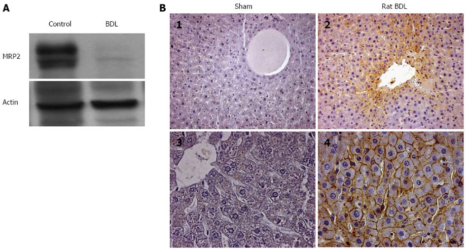
Figure 6 GLT-1/EAAT2 transporter expression in rat liver after bile duct ligation.
A: Immunoblot analysis of multidrug resistance protein 2 transporter and actin protein expression in the liver of sham-operated (control, n = 4) and bile duct ligated (BDL, n = 4) rats. Fifty µg of total protein extract were loaded and subjected to sodium dodecyl sulfate-polyacrylamide gel electrophoresis. Immunoblots are representative of at least three independent experiments; B: Immunohistochemical localization of the GLT-1/EAAT2 transporter in liver sections of sham operated (B1, B3) and bile duct ligated (B2, B4) rats. Immunohistochemistry was performed on paraffin-embedded liver sections using specific antibody for GLT-1 as described in Methods. (B1 and B3) in sham-operated liver rats, no GLT-1 immunoreactivity is observed in hepatocytes. After BDL, (B2 and B4) GLT-1 immunoreactivity is detected as a specific cell surface staining in hepatocytes [original magnification × 200 (B1, B2) and × 400 (B3, B4) respectively].
- Citation: Najimi M, Stéphenne X, Sempoux C, Sokal E. Regulation of hepatic EAAT-2 glutamate transporter expression in human liver cholestasis. World J Gastroenterol 2014; 20(6): 1554-1564
- URL: https://www.wjgnet.com/1007-9327/full/v20/i6/1554.htm
- DOI: https://dx.doi.org/10.3748/wjg.v20.i6.1554


