Copyright
©2014 Baishideng Publishing Group Co.
World J Gastroenterol. Feb 14, 2014; 20(6): 1402-1423
Published online Feb 14, 2014. doi: 10.3748/wjg.v20.i6.1402
Published online Feb 14, 2014. doi: 10.3748/wjg.v20.i6.1402
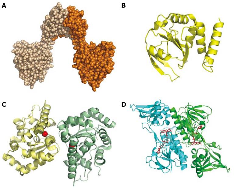
Figure 5 Redox proteins.
A: Space-filling model of the dimer of DsbG (HP0231; PDB 3TDG); B: Cartoon of DsbC (HP0377; Coordinates PDB 4FYC), an enzyme with a thioredoxin-like fold possibly involved in cytochrome c assembly; C: Cartoon of the dimeric Fe-superoxide dismutase (Coordinates PDB 3CEI). The iron ion is represented by a red sphere; D: Dimeric thioredoxin reductase (Coordinates PDB 3ISH). The FAD bound is shown as a ball-and-stick model.
-
Citation: Zanotti G, Cendron L. Structural and functional aspects of the
Helicobacter pylori secretome. World J Gastroenterol 2014; 20(6): 1402-1423 - URL: https://www.wjgnet.com/1007-9327/full/v20/i6/1402.htm
- DOI: https://dx.doi.org/10.3748/wjg.v20.i6.1402


