Copyright
©2014 Baishideng Publishing Group Co.
World J Gastroenterol. Feb 7, 2014; 20(5): 1365-1370
Published online Feb 7, 2014. doi: 10.3748/wjg.v20.i5.1365
Published online Feb 7, 2014. doi: 10.3748/wjg.v20.i5.1365
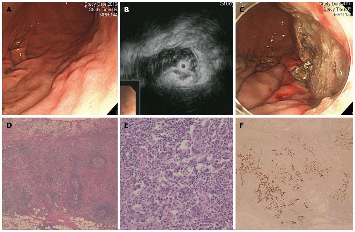
Figure 3 Endoscopic and pathological features of early lymphoepithelioma-like gastric carcinoma (mimicking a subepithelial tumor) treated by endoscopic submucosal dissection (Case 3).
A: White light endoscopy image shows early lymphoepithelioma-like gastric carcinoma (LELC) mimicking a subepithelial tumor; B: Endoscopic ultrasound image shows a homogeneous hypoechoic lesion growing from the muscularis mucosa. Invasion of this lesion into the thickened submucosa layer can also be seen; C: Appearance of the iatrogenic ulcer after endoscopic submucosal dissection; D: Photomicrograph of LELC shows a dense infiltration of lymphocytes in the tumor stroma (HE, × 50); E: Nests of tumor cells separated by a dense infiltration of lymphocytes (HE, × 400); F: In situ hybridization of Epstein-Barr virus-encoded RNA in carcinoma cells (× 50).
- Citation: Lee JY, Kim KM, Min BH, Lee JH, Rhee PL, Kim JJ. Epstein-Barr virus-associated lymphoepithelioma-like early gastric carcinomas and endoscopic submucosal dissection: Case series. World J Gastroenterol 2014; 20(5): 1365-1370
- URL: https://www.wjgnet.com/1007-9327/full/v20/i5/1365.htm
- DOI: https://dx.doi.org/10.3748/wjg.v20.i5.1365


