Copyright
©2014 Baishideng Publishing Group Co.
World J Gastroenterol. Feb 7, 2014; 20(5): 1365-1370
Published online Feb 7, 2014. doi: 10.3748/wjg.v20.i5.1365
Published online Feb 7, 2014. doi: 10.3748/wjg.v20.i5.1365
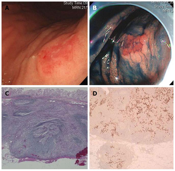
Figure 1 Endoscopic and pathological features of early lymphoepithelioma-like gastric carcinoma treated by endoscopic submucosal dissection (Case 1).
A: White light endoscopy image shows early lymphoepithelioma-like gastric carcinoma (LELC) with surface hyperemia; B: Image of the same sample after treatment with indigocarmine dye; C: Photomicrograph of LELC shows dense lymphocytic infiltration and a pushing margin (HE, × 50); D: Photomicrograph of Epstein-Barr virus-encoded RNA detected by in situ hybridization (× 50). Note the positive staining localized specifically to the nuclei of tumor cells, while no signal can be seen in the lymphocytes.
- Citation: Lee JY, Kim KM, Min BH, Lee JH, Rhee PL, Kim JJ. Epstein-Barr virus-associated lymphoepithelioma-like early gastric carcinomas and endoscopic submucosal dissection: Case series. World J Gastroenterol 2014; 20(5): 1365-1370
- URL: https://www.wjgnet.com/1007-9327/full/v20/i5/1365.htm
- DOI: https://dx.doi.org/10.3748/wjg.v20.i5.1365


