Copyright
©2014 Baishideng Publishing Group Co.
World J Gastroenterol. Feb 7, 2014; 20(5): 1180-1191
Published online Feb 7, 2014. doi: 10.3748/wjg.v20.i5.1180
Published online Feb 7, 2014. doi: 10.3748/wjg.v20.i5.1180
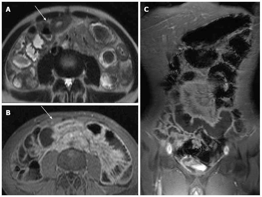
Figure 15 Magnetic resonance enterography allows an overview about involved bowel segments.
In this case there was an isolated involvement of the jejunum. A: Axial T2w sequence showing thickened bowel wall without edema; B, C: Axial and coronal fat saturated contrast-enhanced T1w sequence demonstrating transmural enhancement in Crohn’s disease. T1w: T1 weighted; T2w: T2 weighted.
- Citation: Mentzel HJ, Reinsch S, Kurzai M, Stenzel M. Magnetic resonance imaging in children and adolescents with chronic inflammatory bowel disease. World J Gastroenterol 2014; 20(5): 1180-1191
- URL: https://www.wjgnet.com/1007-9327/full/v20/i5/1180.htm
- DOI: https://dx.doi.org/10.3748/wjg.v20.i5.1180


