Copyright
©2014 Baishideng Publishing Group Co.
World J Gastroenterol. Feb 7, 2014; 20(5): 1180-1191
Published online Feb 7, 2014. doi: 10.3748/wjg.v20.i5.1180
Published online Feb 7, 2014. doi: 10.3748/wjg.v20.i5.1180
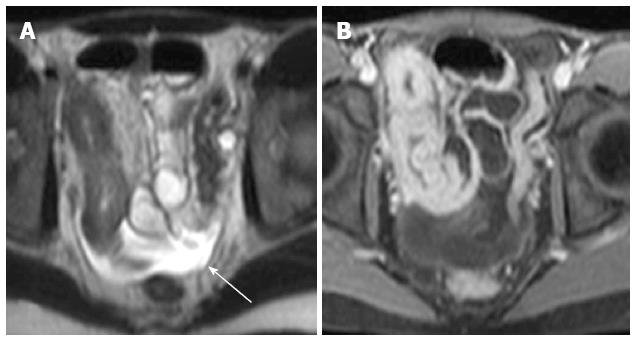
Figure 11 Thirteen years old girl with Crohn’s disease.
A: T2w image demonstrating strong hyperintense ascites. Arrow marks the free ascites; B: T1w sequence after contrast application. Fluid is not as easily detected as in T2w images. T1w: T1 weighted; T2w: T2 weighted.
- Citation: Mentzel HJ, Reinsch S, Kurzai M, Stenzel M. Magnetic resonance imaging in children and adolescents with chronic inflammatory bowel disease. World J Gastroenterol 2014; 20(5): 1180-1191
- URL: https://www.wjgnet.com/1007-9327/full/v20/i5/1180.htm
- DOI: https://dx.doi.org/10.3748/wjg.v20.i5.1180


