Copyright
©2014 Baishideng Publishing Group Inc.
World J Gastroenterol. Dec 28, 2014; 20(48): 18495-18502
Published online Dec 28, 2014. doi: 10.3748/wjg.v20.i48.18495
Published online Dec 28, 2014. doi: 10.3748/wjg.v20.i48.18495
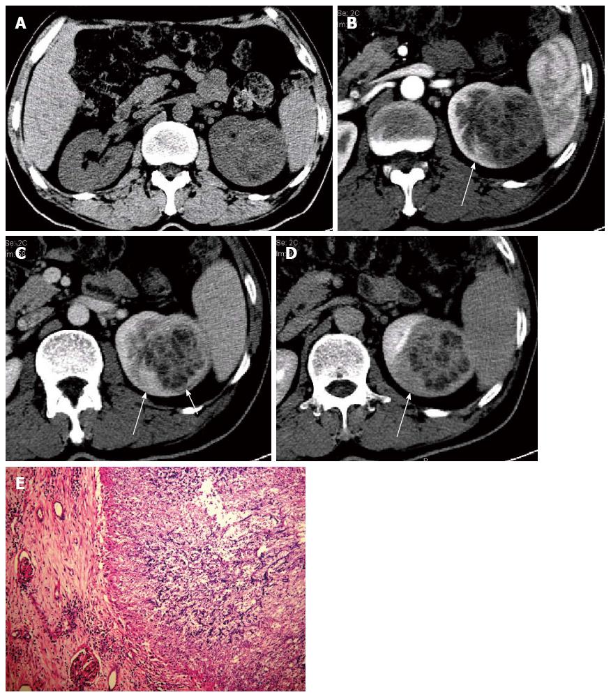
Figure 1 Renal aspergillosis in a 47-year-old allogenetic orthotropic liver transplantation recipient (Case one).
A: Unenhanced transverse CT image reveals a large inhomogeneous hypo-attenuation focus in the left kidney; B: Transverse image in the contrast-enhanced cortical nephrographic phase; C: Transverse image in the parenchymal nephrographic phase; D: Transverse image in the excretory phase. The focus shows progressively enhanced abscess walls and separations with unenhanced necrotic areas. A wedge-shaped relatively lowly enhanced area (long arrow) is detected around the focus in B, C and D, which presents with a gradually increased density difference with the surrounding parenchyma and is confirmed to be the secondary change to chronic ischemia by histopathology. A relatively lowly enhanced thin layer (short arrow) is clearly detected in the parenchymal nephrographic phase, which is dense fibrous connective tissues in the abscess wall; E: Pathological findings under a light microscope (HE, × 100), which shows the abscesses with necrosis and amounts of aspergillus hyphae, spores and neutrophils. CT: Computed tomography; HE: Hematoxylin-eosin.
- Citation: Meng XC, Jiang T, Yi SH, Xie PY, Guo YF, Quan L, Zhou J, Zhu KS, Shan H. Renal aspergillosis after liver transplantation: Clinical and imaging manifestations in two cases. World J Gastroenterol 2014; 20(48): 18495-18502
- URL: https://www.wjgnet.com/1007-9327/full/v20/i48/18495.htm
- DOI: https://dx.doi.org/10.3748/wjg.v20.i48.18495


