Copyright
©2014 Baishideng Publishing Group Inc.
World J Gastroenterol. Dec 28, 2014; 20(48): 18487-18494
Published online Dec 28, 2014. doi: 10.3748/wjg.v20.i48.18487
Published online Dec 28, 2014. doi: 10.3748/wjg.v20.i48.18487
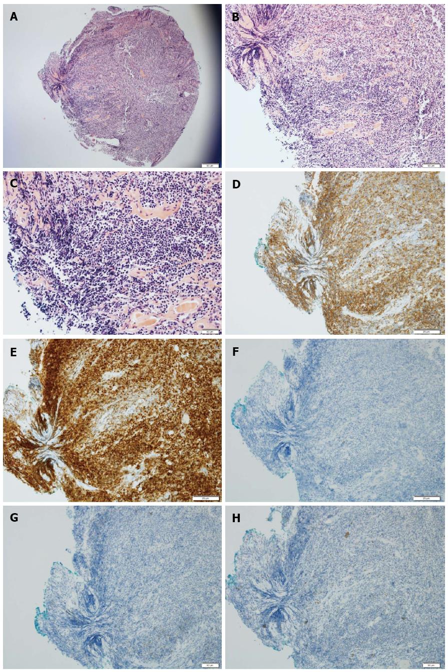
Figure 2 Histopathologic findings.
A-C: Hematoxylin-eosin staining of the biopsy specimen showing dense proliferation of atypical lymphoid cells and infiltrated centrocyte-like cells in the mucosal layer (magnification: A, 40 ×; B, 100 ×; C, 200 ×); D: CD20-positive; E: Bcl-2-positive; F: CD10-negative; G: CD23-negative; H: Bcl-6-negative immunoreactivity of the biopsy specimen (100 × magnification).
- Citation: Seo SW, Lee SH, Lee DJ, Kim KM, Kang JK, Kim DW, Lee JH. Colonic mucosa-associated lymphoid tissue lymphoma identified by chromoendoscopy. World J Gastroenterol 2014; 20(48): 18487-18494
- URL: https://www.wjgnet.com/1007-9327/full/v20/i48/18487.htm
- DOI: https://dx.doi.org/10.3748/wjg.v20.i48.18487


