Copyright
©2014 Baishideng Publishing Group Inc.
World J Gastroenterol. Dec 28, 2014; 20(48): 18480-18486
Published online Dec 28, 2014. doi: 10.3748/wjg.v20.i48.18480
Published online Dec 28, 2014. doi: 10.3748/wjg.v20.i48.18480
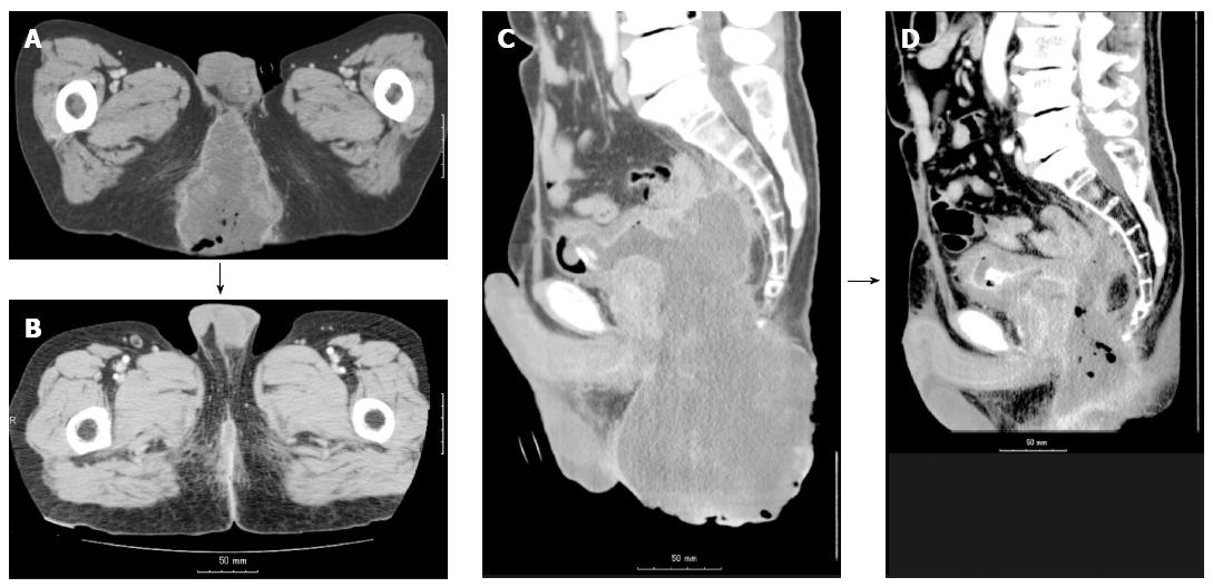
Figure 2 Computed tomography image.
A: An axial computed tomography (CT) image before radiotherapy. A massive tumor is seen in the perineum, and a part of the tumor is protruding. Both lower extremities are elevated because the patient could not stretch both legs. B: An axial CT image on day 120 from the start of the radiotherapy. The macroscopic tumor disappeared, and the patient could stretch both lower extremities; C: A sagittal CT image before the radiotherapy; D: A part of the bladder wall has been lost to invasion of the massive solid tumor.
- Citation: Nomiya T, Akamatsu H, Harada M, Ota I, Hagiwara Y, Ichikawa M, Miwa M, Kawashiro S, Hagiwara M, Chin M, Hashizume E, Nemoto K. Modified simultaneous integrated boost radiotherapy for an unresectable huge refractory pelvic tumor diagnosed as a rectal adenocarcinoma. World J Gastroenterol 2014; 20(48): 18480-18486
- URL: https://www.wjgnet.com/1007-9327/full/v20/i48/18480.htm
- DOI: https://dx.doi.org/10.3748/wjg.v20.i48.18480


