Copyright
©2014 Baishideng Publishing Group Inc.
World J Gastroenterol. Dec 28, 2014; 20(48): 18375-18383
Published online Dec 28, 2014. doi: 10.3748/wjg.v20.i48.18375
Published online Dec 28, 2014. doi: 10.3748/wjg.v20.i48.18375
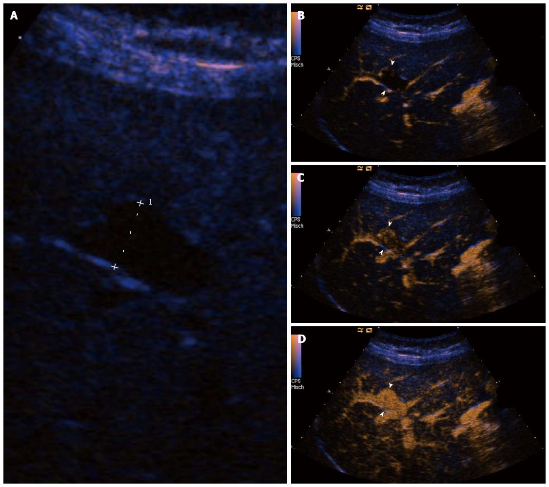
Figure 4 Patient 5: Portal venous system aneurysm in a 58-year-old man who was referred because of elevated liver function tests.
A: Right transverse subcostal view shows a focal saccular dilatation of the right main branch of the portal vein, with a maximum diameter of 25 mm (caliper 1); B-D: Right transverse subcostal view shows the contrast-enhanced ultrasound characteristics of the portal venous system aneurysm (PVSA), which is not enhanced in the arterial phase (16 s, arrowheads, B) and starts to enhance through the early portal venous phase (18 s, arrowheads, C). The PVSA is completely enhanced during portal venous phase (21 s, arrowheads, D).
- Citation: Tana C, Dietrich CF, Badea R, Chiorean L, Carrieri V, Schiavone C. Contrast-enhanced ultrasound in portal venous system aneurysms: A multi-center study. World J Gastroenterol 2014; 20(48): 18375-18383
- URL: https://www.wjgnet.com/1007-9327/full/v20/i48/18375.htm
- DOI: https://dx.doi.org/10.3748/wjg.v20.i48.18375


