Copyright
©2014 Baishideng Publishing Group Inc.
World J Gastroenterol. Dec 28, 2014; 20(48): 18375-18383
Published online Dec 28, 2014. doi: 10.3748/wjg.v20.i48.18375
Published online Dec 28, 2014. doi: 10.3748/wjg.v20.i48.18375
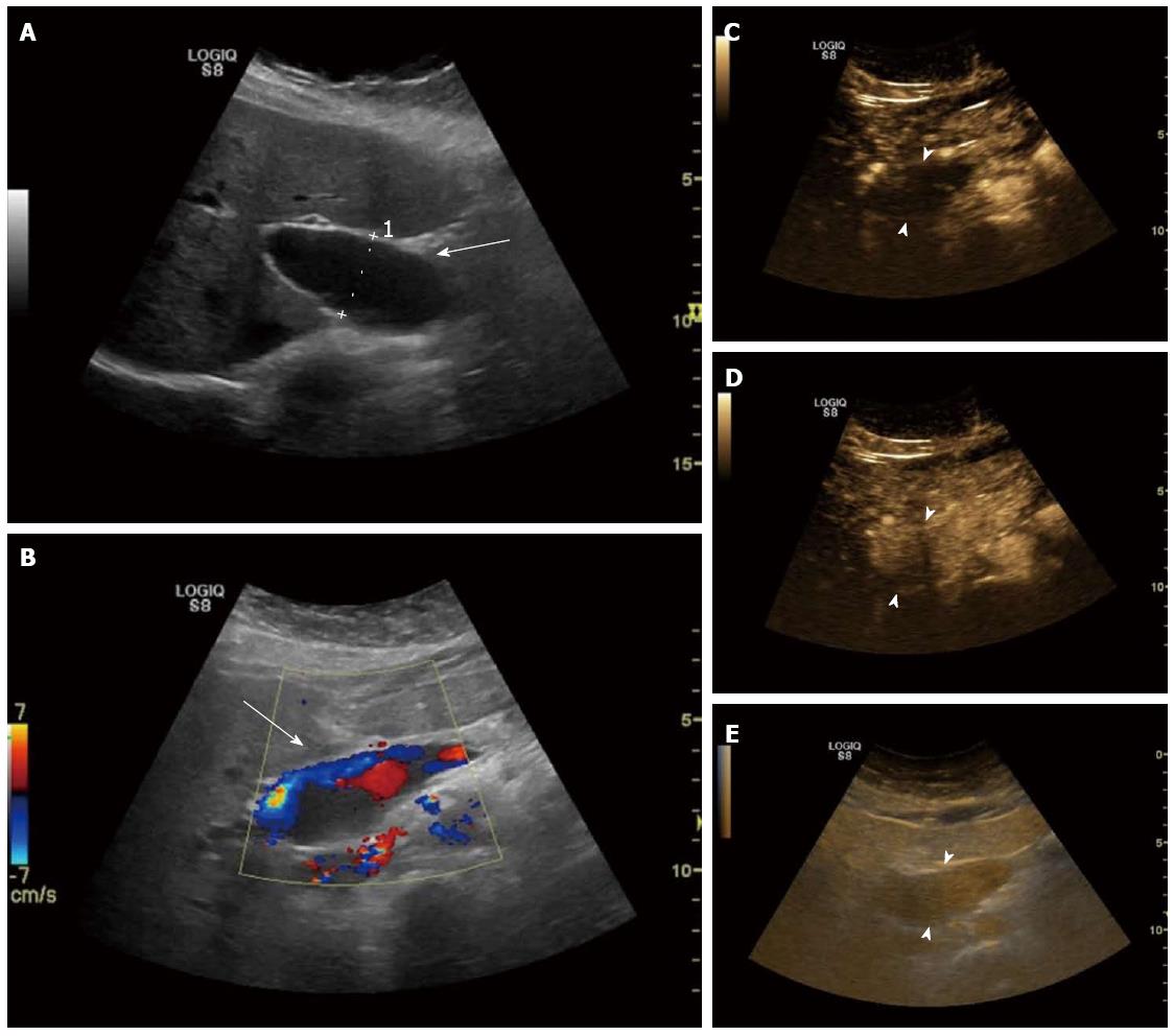
Figure 1 Patient 1: Portal venous system aneurysm in a 56-year-old woman with a history of overweight and dyslipidemia, who was referred because of suspected nonalcoholic fatty liver disease.
A: Right oblique sagittal view shows a focal fusiform dilatation of the extrahepatic segment of the main portal vein (arrow), with a maximum diameter of 30 mm (caliper 1); B: Right oblique sagittal view with color Doppler shows “to-and-fro” flow signal inside the lesion (arrow); C: Right oblique sagittal view shows the contrast-enhanced ultrasound characteristics of the portal venous system aneurysm which is not enhanced in the arterial phase (17 s, arrowheads); D: Homogenously enhanced throughout the early portal venous phase (22 s, arrowheads); E: Persistently enhanced with slow washout in the late phase (301 s, arrowheads).
- Citation: Tana C, Dietrich CF, Badea R, Chiorean L, Carrieri V, Schiavone C. Contrast-enhanced ultrasound in portal venous system aneurysms: A multi-center study. World J Gastroenterol 2014; 20(48): 18375-18383
- URL: https://www.wjgnet.com/1007-9327/full/v20/i48/18375.htm
- DOI: https://dx.doi.org/10.3748/wjg.v20.i48.18375


