Copyright
©2014 Baishideng Publishing Group Inc.
World J Gastroenterol. Dec 21, 2014; 20(47): 17962-17969
Published online Dec 21, 2014. doi: 10.3748/wjg.v20.i47.17962
Published online Dec 21, 2014. doi: 10.3748/wjg.v20.i47.17962
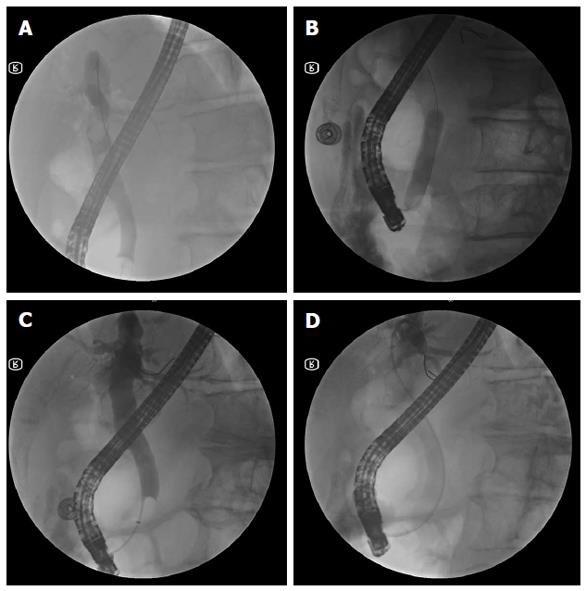
Figure 1 Fluoroscopic view of large-balloon dilatation following limited sphincterotomy.
A: Cholangiogram demonstrating two large stones within the dilated bile duct; B: A large balloon inflated across the papilla over the guidewire; C: The cholangiogram following complete stone removal showed no residual filling defect in the bile duct; D: The placement of a nasobiliary drainage tube.
-
Citation: Guo SB, Meng H, Duan ZJ, Li CY. Small sphincterotomy combined with endoscopic papillary large balloon dilation
vs sphincterotomy alone for removal of common bile duct stones. World J Gastroenterol 2014; 20(47): 17962-17969 - URL: https://www.wjgnet.com/1007-9327/full/v20/i47/17962.htm
- DOI: https://dx.doi.org/10.3748/wjg.v20.i47.17962


