Copyright
©2014 Baishideng Publishing Group Inc.
World J Gastroenterol. Dec 21, 2014; 20(47): 17955-17961
Published online Dec 21, 2014. doi: 10.3748/wjg.v20.i47.17955
Published online Dec 21, 2014. doi: 10.3748/wjg.v20.i47.17955
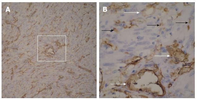
Figure 6 Representative findings from immunohistochemical examination of small bowel gastrointestinal stromal tumor.
A: CD34-positive cells, as well as numerous blood vessels of various diameters (magnification × 100), are shown; B: The magnified imaging (A, white box) in which the CD34-positive tumor cells (B, black arrows) and vascular endothelial cells (B, white arrows) are clearly evident (magnification × 400).
- Citation: Chen YT, Sun HL, Luo JH, Ni JY, Chen D, Jiang XY, Zhou JX, Xu LF. Interventional digital subtraction angiography for small bowel gastrointestinal stromal tumors with bleeding. World J Gastroenterol 2014; 20(47): 17955-17961
- URL: https://www.wjgnet.com/1007-9327/full/v20/i47/17955.htm
- DOI: https://dx.doi.org/10.3748/wjg.v20.i47.17955


