Copyright
©2014 Baishideng Publishing Group Inc.
World J Gastroenterol. Dec 21, 2014; 20(47): 17955-17961
Published online Dec 21, 2014. doi: 10.3748/wjg.v20.i47.17955
Published online Dec 21, 2014. doi: 10.3748/wjg.v20.i47.17955
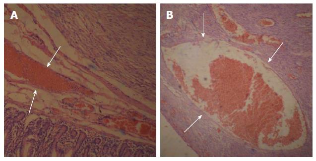
Figure 5 Representative findings from pathological examination of small bowel gastrointestinal stromal tumor.
Two microscopic fields (A, B) of a hematoxylin-eosin stained gastrointestinal stromal tumor (GIST) shows a rich variety of tumor vessels filled with red blood cells (white arrows) (magnification × 40).
- Citation: Chen YT, Sun HL, Luo JH, Ni JY, Chen D, Jiang XY, Zhou JX, Xu LF. Interventional digital subtraction angiography for small bowel gastrointestinal stromal tumors with bleeding. World J Gastroenterol 2014; 20(47): 17955-17961
- URL: https://www.wjgnet.com/1007-9327/full/v20/i47/17955.htm
- DOI: https://dx.doi.org/10.3748/wjg.v20.i47.17955


