Copyright
©2014 Baishideng Publishing Group Inc.
World J Gastroenterol. Dec 21, 2014; 20(47): 17746-17755
Published online Dec 21, 2014. doi: 10.3748/wjg.v20.i47.17746
Published online Dec 21, 2014. doi: 10.3748/wjg.v20.i47.17746
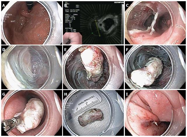
Figure 5 Submucosal tunneling endoscopic resection technique.
A: Gastroesophageal junction lesion seen on retroflexion during endoscopy; B: Endoscopic ultrasound probe demonstrates hypoechoic muscularis propria based lesion; C: Creation of submucosal tunnel parallel to esophageal lumen; D: Endoscopic submucosal dissection (ESD) with submucosal tunnel; E-F: Freeing of submucosal lesion via ESD; G: Removal of submucosal lesion from tunnel with biopsy forceps; H: 2.5 cm leiomyoma; I: Endoscopic sutured closure of mucosal entrance to tunnel.
- Citation: Friedel D, Modayil R, Stavropoulos SN. Per-oral endoscopic myotomy: Major advance in achalasia treatment and in endoscopic surgery. World J Gastroenterol 2014; 20(47): 17746-17755
- URL: https://www.wjgnet.com/1007-9327/full/v20/i47/17746.htm
- DOI: https://dx.doi.org/10.3748/wjg.v20.i47.17746


