Copyright
©2014 Baishideng Publishing Group Inc.
World J Gastroenterol. Dec 14, 2014; 20(46): 17680-17685
Published online Dec 14, 2014. doi: 10.3748/wjg.v20.i46.17680
Published online Dec 14, 2014. doi: 10.3748/wjg.v20.i46.17680
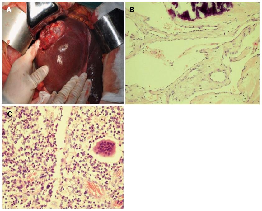
Figure 3 Operative exploration finding and histology.
A: Sclerotic liver cavernous hemangioma was located in the atrophic right lobe but the left lobe was remarkably hyperplastic with the displacement of the hilus towards the right side; B: Histological staining showed a pathology of liver cavernous hemangioma [hematoxylin-eosin (HE, × 200); C: Sclerosing cholangitis with complicating chronic pyogenic inflammation (HE, × 200).
- Citation: Jin S, Shi XJ, Sun XD, Wang SY, Wang GY. Sclerosing cholangitis secondary to bleomycin-iodinated embolization for liver hemangioma. World J Gastroenterol 2014; 20(46): 17680-17685
- URL: https://www.wjgnet.com/1007-9327/full/v20/i46/17680.htm
- DOI: https://dx.doi.org/10.3748/wjg.v20.i46.17680


