Copyright
©2014 Baishideng Publishing Group Inc.
World J Gastroenterol. Dec 14, 2014; 20(46): 17680-17685
Published online Dec 14, 2014. doi: 10.3748/wjg.v20.i46.17680
Published online Dec 14, 2014. doi: 10.3748/wjg.v20.i46.17680
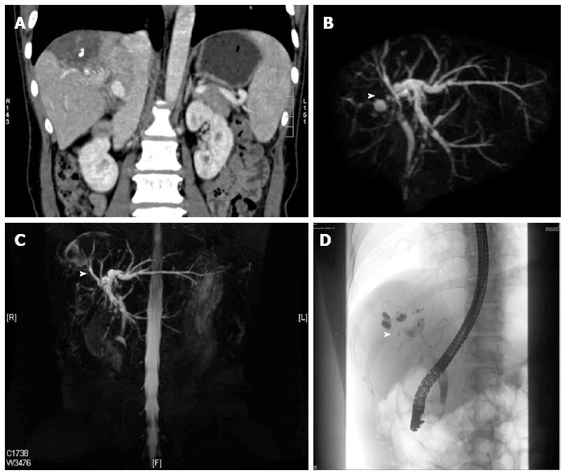
Figure 2 Three-dimensional reconstructed computed tomography, magnetic retrograde cholangio- pancreatography and endoscopic retrograde cholangio- pancreatography.
A: Liver three-dimensional (3D)-computed tomography scan; B, C: 3D-magnetic retrograde cholangio- pancreatography scan confirms the dilation of the left hepatic duct and the narrowing of the right hepatic duct (the white arrowheads indicate a soft tissue-like mass located at the convergence of the left and right hepatic ducts); D: Endoscopic retrograde cholangio- pancreatography shows no visualization of the hilar bile duct and poor visualization of the intrahepatic bile ducts (white arrowhead).
- Citation: Jin S, Shi XJ, Sun XD, Wang SY, Wang GY. Sclerosing cholangitis secondary to bleomycin-iodinated embolization for liver hemangioma. World J Gastroenterol 2014; 20(46): 17680-17685
- URL: https://www.wjgnet.com/1007-9327/full/v20/i46/17680.htm
- DOI: https://dx.doi.org/10.3748/wjg.v20.i46.17680


