Copyright
©2014 Baishideng Publishing Group Inc.
World J Gastroenterol. Dec 14, 2014; 20(46): 17674-17679
Published online Dec 14, 2014. doi: 10.3748/wjg.v20.i46.17674
Published online Dec 14, 2014. doi: 10.3748/wjg.v20.i46.17674
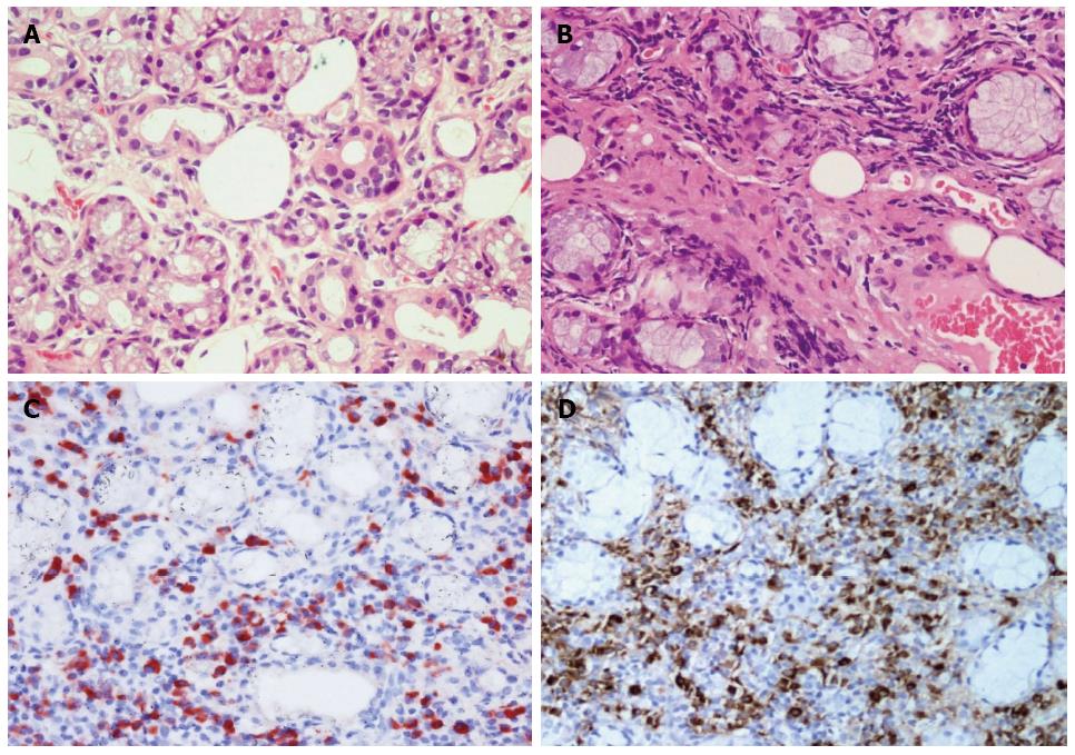
Figure 2 Histological findings of the excisional biopsy specimen of the lower lip lumps.
Hematoxylin and eosin (HE) stain showing normal labial gland (A, × 400); acinar atrophy and lymphoplasmacyte infiltration and fibrosis from the patient (B, × 400); immunohistochemical staining for IgG4 (red color) (C, × 400) or IgG (yellow color) (D, × 400) in plasma cells from the patient.
- Citation: Sun L, Zhou Q, Brigstock DR, Yan S, Xiu M, Piao RL, Gao YH, Gao RP. Focal autoimmune pancreatitis and chronic sclerosing sialadenitis mimicking pancreatic cancer and neck metastasis. World J Gastroenterol 2014; 20(46): 17674-17679
- URL: https://www.wjgnet.com/1007-9327/full/v20/i46/17674.htm
- DOI: https://dx.doi.org/10.3748/wjg.v20.i46.17674


