Copyright
©2014 Baishideng Publishing Group Inc.
World J Gastroenterol. Dec 14, 2014; 20(46): 17666-17669
Published online Dec 14, 2014. doi: 10.3748/wjg.v20.i46.17666
Published online Dec 14, 2014. doi: 10.3748/wjg.v20.i46.17666
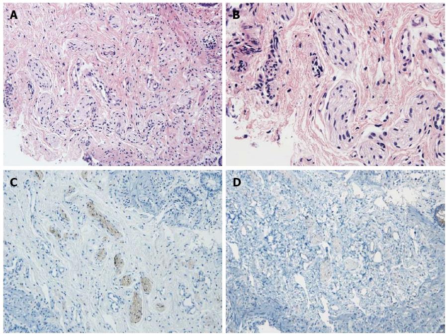
Figure 2 Histological and immunohistochemical features of the rectal biopsy specimen.
A: Increased numbers of nerve bundles can be seen in the submucosal layer (HE, × 200); B: High-powered magnification of the submucosal nerve plexus shows no ganglion cells (HE, × 400). Immunohistochemically; S-100 (× 200) (C) and bcl-2 (× 200) (D) staining show no ganglion cells in the submucosal nerves. HE: Hematoxylin-eosin staining.
- Citation: Park HW, Cho MJ, Kim WY, Kwak BO, Kim MH. Hirschsprung’s disease in twin to twin transfusion syndrome: A case report. World J Gastroenterol 2014; 20(46): 17666-17669
- URL: https://www.wjgnet.com/1007-9327/full/v20/i46/17666.htm
- DOI: https://dx.doi.org/10.3748/wjg.v20.i46.17666


