Copyright
©2014 Baishideng Publishing Group Inc.
World J Gastroenterol. Dec 14, 2014; 20(46): 17666-17669
Published online Dec 14, 2014. doi: 10.3748/wjg.v20.i46.17666
Published online Dec 14, 2014. doi: 10.3748/wjg.v20.i46.17666
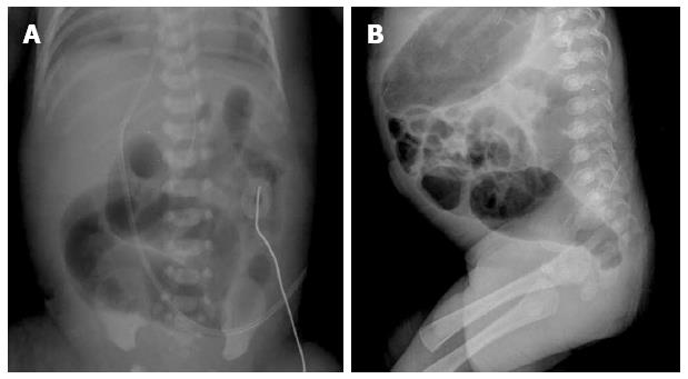
Figure 1 Plain X-ray of the abdomen and pelvis.
A: X-ray of the abdomen showing marked dilatation of the sigmoid colon on 4 d; B: Lateral pelvic film showing dilatation of the sigmoid colon and a narrowed transition zone with a reversed recto-sigmoid ratio on 59 d.
- Citation: Park HW, Cho MJ, Kim WY, Kwak BO, Kim MH. Hirschsprung’s disease in twin to twin transfusion syndrome: A case report. World J Gastroenterol 2014; 20(46): 17666-17669
- URL: https://www.wjgnet.com/1007-9327/full/v20/i46/17666.htm
- DOI: https://dx.doi.org/10.3748/wjg.v20.i46.17666


