Copyright
©2014 Baishideng Publishing Group Inc.
World J Gastroenterol. Dec 14, 2014; 20(46): 17661-17665
Published online Dec 14, 2014. doi: 10.3748/wjg.v20.i46.17661
Published online Dec 14, 2014. doi: 10.3748/wjg.v20.i46.17661
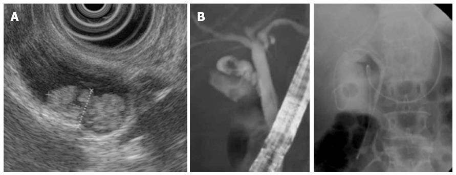
Figure 2 Endoscopic ultrasound images.
A: Endoscopic ultrasound revealed a hyperechoic papillary mass, measuring 16 mm × 8 mm in diameter, in the neck of the gallbladder, as well as multiple small polyps; B: Endoscopic naso-gallbladder drainage was performed to enable cytological examination of the bile within the gallbladder.
- Citation: Mori Y, Sato N, Matayoshi N, Tamura T, Minagawa N, Shibao K, Higure A, Nakamoto M, Taguchi M, Yamaguchi K. Rare combination of familial adenomatous polyposis and gallbladder polyps. World J Gastroenterol 2014; 20(46): 17661-17665
- URL: https://www.wjgnet.com/1007-9327/full/v20/i46/17661.htm
- DOI: https://dx.doi.org/10.3748/wjg.v20.i46.17661


