Copyright
©2014 Baishideng Publishing Group Inc.
World J Gastroenterol. Dec 14, 2014; 20(46): 17661-17665
Published online Dec 14, 2014. doi: 10.3748/wjg.v20.i46.17661
Published online Dec 14, 2014. doi: 10.3748/wjg.v20.i46.17661
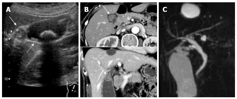
Figure 1 Diagnostic imaging results.
A: Abdominal ultrasound showing an isoechoic mass in the neck of the gallbladder and multiple small polyps measuring 3-16 mm in diameter (arrows) throughout the gallbladder (a 22 mm gallbladder stone was also identified); B: CT showed a contrast-enhanced mass (arrows) in the neck of the gallbladder; C: Magnetic resonance cholangiopancreatography showed no other abnormalities of the biliary and pancreatic ductal systems.
- Citation: Mori Y, Sato N, Matayoshi N, Tamura T, Minagawa N, Shibao K, Higure A, Nakamoto M, Taguchi M, Yamaguchi K. Rare combination of familial adenomatous polyposis and gallbladder polyps. World J Gastroenterol 2014; 20(46): 17661-17665
- URL: https://www.wjgnet.com/1007-9327/full/v20/i46/17661.htm
- DOI: https://dx.doi.org/10.3748/wjg.v20.i46.17661


