Copyright
©2014 Baishideng Publishing Group Inc.
World J Gastroenterol. Dec 14, 2014; 20(46): 17541-17551
Published online Dec 14, 2014. doi: 10.3748/wjg.v20.i46.17541
Published online Dec 14, 2014. doi: 10.3748/wjg.v20.i46.17541
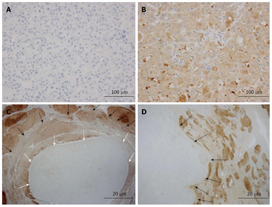
Figure 1 Immunohistochemical staining of liver fatty acid-binding protein in hepatocellular carcinoma.
A: Representative liver fatty acid-binding protein (L-FABP)-negative case of hepatocellular carcinoma (HCC) identified by examination using tissue microarrays; B: Representative L-FABP-positive case of HCC identified by examination using tissue microarrays; C: Focal downregulation of L-FABP expression was observed in a case of small HCC (white arrows show the boundary between L-FABP-negative and L-FABP-positive areas, and black arrows show the boundary between tumorous and non-tumorous areas); D: Diffuse downregulation was observed in a case of large HCC (black arrows show the boundary between tumorous and non-tumorous areas).
- Citation: Inoue M, Takahashi Y, Fujii T, Kitagawa M, Fukusato T. Significance of downregulation of liver fatty acid-binding protein in hepatocellular carcinoma. World J Gastroenterol 2014; 20(46): 17541-17551
- URL: https://www.wjgnet.com/1007-9327/full/v20/i46/17541.htm
- DOI: https://dx.doi.org/10.3748/wjg.v20.i46.17541


