Copyright
©2014 Baishideng Publishing Group Inc.
World J Gastroenterol. Dec 14, 2014; 20(46): 17407-17415
Published online Dec 14, 2014. doi: 10.3748/wjg.v20.i46.17407
Published online Dec 14, 2014. doi: 10.3748/wjg.v20.i46.17407
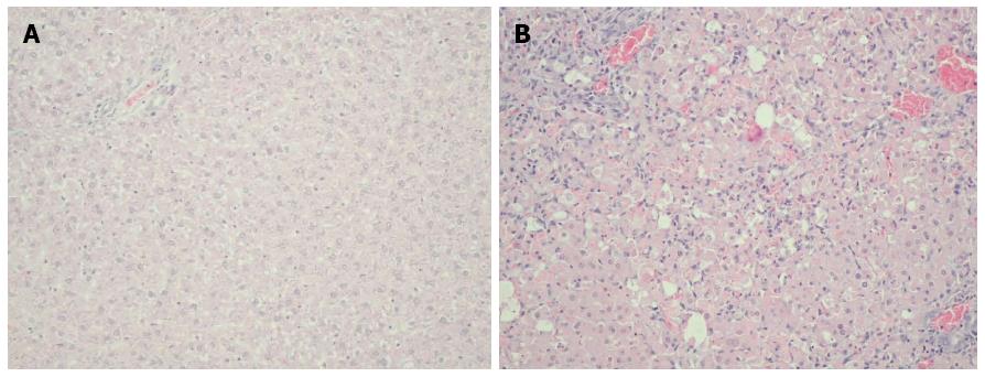
Figure 1 Histopathological image of the liver.
A: Sham rats. Lack of morphological changes in the liver from sham Sprague-Dawley rats (Group 1) 48 h after saline injection. Hematoxylin and eosin staining, light microscopy, magnification × 20; B: Test rats. Massive necrosis of hepatocytes in the liver of Sprague-Dawley rats (Group 2) 48 h after galactosamine injection. Hematoxylin and eosin staining, light microscopy, magnification × 20.
- Citation: Saracyn M, Brytan M, Zdanowski R, Ząbkowski T, Dyrla P, Patera J, Wojtuń S, Kozłowski W, Wańkowicz Z. Hepatoprotective effect of nitric oxide in experimental model of acute hepatic failure. World J Gastroenterol 2014; 20(46): 17407-17415
- URL: https://www.wjgnet.com/1007-9327/full/v20/i46/17407.htm
- DOI: https://dx.doi.org/10.3748/wjg.v20.i46.17407


