Copyright
©2014 Baishideng Publishing Group Inc.
World J Gastroenterol. Dec 14, 2014; 20(46): 17399-17406
Published online Dec 14, 2014. doi: 10.3748/wjg.v20.i46.17399
Published online Dec 14, 2014. doi: 10.3748/wjg.v20.i46.17399
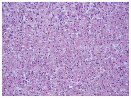
Figure 4 Light microscopy of a liver biopsy.
After the passing of the animal subjects, liver tissues were prepared and stained for hematoxylin and eosin. Hepatocytes showed diffused swelling, were sinusoidal, and became narrow after pressure. The cytoplasm of hepatocytes appeared loose, and there were signs of vacuolar degeneration, nuclear fragmentation and dissolution. Part of the portal area showed a small amount of neutrophils and lymphocytes (× 200).
- Citation: Zhang Z, Zhao YC, Cheng Y, Jian GD, Pan MX, Gao Y. Hybrid bioartificial liver support in cynomolgus monkeys with D-galactosamine-induced acute liver failure. World J Gastroenterol 2014; 20(46): 17399-17406
- URL: https://www.wjgnet.com/1007-9327/full/v20/i46/17399.htm
- DOI: https://dx.doi.org/10.3748/wjg.v20.i46.17399


