Copyright
©2014 Baishideng Publishing Group Inc.
World J Gastroenterol. Dec 7, 2014; 20(45): 17247-17253
Published online Dec 7, 2014. doi: 10.3748/wjg.v20.i45.17247
Published online Dec 7, 2014. doi: 10.3748/wjg.v20.i45.17247
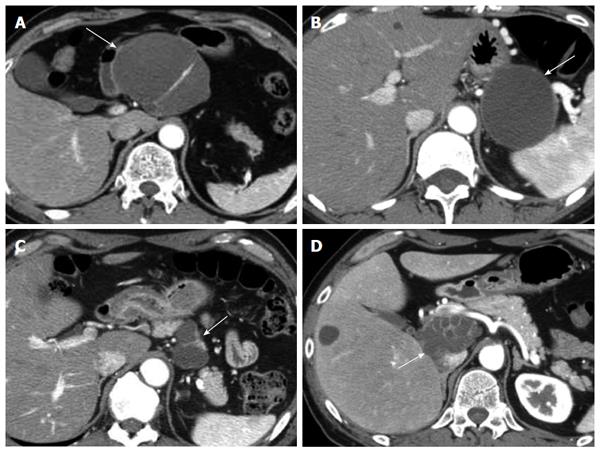
Figure 2 Computed tomography findings.
All lesions were well-defined and were exophytic off the pancreatic parenchyma. The wall and septum of the cysts were enhanced. A: Findings of Patient #1, the lesion was localized in the body of the pancreas and had a multilocular cystic appearance (arrow); B: Findings of Patient #2, the lesion was localized in the tail of the pancreas and had a unilocular cystic appearance (arrow); C: Findings of Patient #3, the lesion was localized in the body of the pancreas and had a multilocular cystic appearance (arrow); D: Findings of Patient #4, the lesion was localized in the head of the pancreas and had a multilocular cystic appearance (arrow).
- Citation: Terakawa H, Makino I, Nakagawara H, Miyashita T, Tajima H, Kitagawa H, Fujimura T, Inoue D, Kozaka K, Gabata T, Ohta T. Clinical and radiological feature of lymphoepithelial cyst of the pancreas. World J Gastroenterol 2014; 20(45): 17247-17253
- URL: https://www.wjgnet.com/1007-9327/full/v20/i45/17247.htm
- DOI: https://dx.doi.org/10.3748/wjg.v20.i45.17247


