Copyright
©2014 Baishideng Publishing Group Inc.
World J Gastroenterol. Dec 7, 2014; 20(45): 17244-17246
Published online Dec 7, 2014. doi: 10.3748/wjg.v20.i45.17244
Published online Dec 7, 2014. doi: 10.3748/wjg.v20.i45.17244
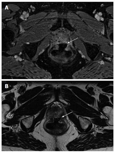
Figure 2 Large ulceration (indicated by white arrow) with regular edges sitting without local infectious complication or digestive fistula on pelvic magnetic resonance imaging performed 1 mo after endoscopic argon plasma coagulationprocedure.
A: Axial T1-weighted section after injection of gadolinium chelate; B: Axial T2-weighted section.
- Citation: Koessler T, Servois V, Mariani P, Aubert E, Cacheux W. Rectal ulcer: Due to ketoprofen, argon plasma coagulation and prostatic brachytherapy. World J Gastroenterol 2014; 20(45): 17244-17246
- URL: https://www.wjgnet.com/1007-9327/full/v20/i45/17244.htm
- DOI: https://dx.doi.org/10.3748/wjg.v20.i45.17244


