Copyright
©2014 Baishideng Publishing Group Inc.
World J Gastroenterol. Dec 7, 2014; 20(45): 17084-17091
Published online Dec 7, 2014. doi: 10.3748/wjg.v20.i45.17084
Published online Dec 7, 2014. doi: 10.3748/wjg.v20.i45.17084
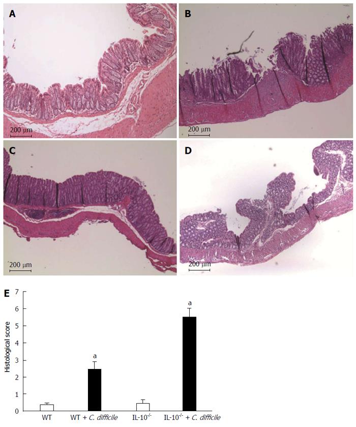
Figure 4 Histopathologic examination of the colonic tissue (× 100).
Hematoxylin and eosin staining of colon tissues from A: Wild type (WT); B: WT challenged with Clostridium difficile (C. difficile); C: Interleukin 10-deficient (IL-10-/-); D: IL-10-/- challenged with C. difficile; E: Quantification of colitis severity. Error bars indicate standard error of the mean; aP < 0.05 vs untreated.
-
Citation: Kim MN, Koh SJ, Kim JM, Im JP, Jung HC, Kim JS.
Clostridium difficile infection aggravates colitis in interleukin 10-deficient mice. World J Gastroenterol 2014; 20(45): 17084-17091 - URL: https://www.wjgnet.com/1007-9327/full/v20/i45/17084.htm
- DOI: https://dx.doi.org/10.3748/wjg.v20.i45.17084


