Copyright
©2014 Baishideng Publishing Group Inc.
World J Gastroenterol. Dec 7, 2014; 20(45): 17037-17048
Published online Dec 7, 2014. doi: 10.3748/wjg.v20.i45.17037
Published online Dec 7, 2014. doi: 10.3748/wjg.v20.i45.17037
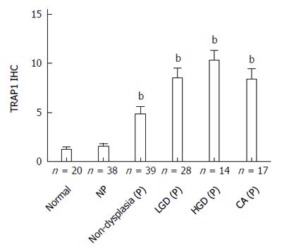
Figure 1 Tumor necrosis factor receptor-associated protein 1 staining increases with ulcerative colitis progression at both non-dysplastic and dysplastic sites.
Plot depicts the average immunohistochemistry (IHC) score ± SE for rectal samples from normal controls, non-progressors, progressors at non-dysplastic rectal sites, and progressors at dysplastic sites. The number of patient is indicated. bP < 0.01 using Mann Whitney test vs normal controls. NP: Non-progressors; P: Progressors; LGD: Low grade dysplasia; HGD: High grade dysplasia; CA: Cancer; TRAP1: Tumor necrosis factor receptor-associated protein 1.
- Citation: Chen R, Pan S, Lai K, Lai LA, Crispin DA, Bronner MP, Brentnall TA. Up-regulation of mitochondrial chaperone TRAP1 in ulcerative colitis associated colorectal cancer. World J Gastroenterol 2014; 20(45): 17037-17048
- URL: https://www.wjgnet.com/1007-9327/full/v20/i45/17037.htm
- DOI: https://dx.doi.org/10.3748/wjg.v20.i45.17037


