Copyright
©2014 Baishideng Publishing Group Inc.
World J Gastroenterol. Dec 7, 2014; 20(45): 16984-16995
Published online Dec 7, 2014. doi: 10.3748/wjg.v20.i45.16984
Published online Dec 7, 2014. doi: 10.3748/wjg.v20.i45.16984
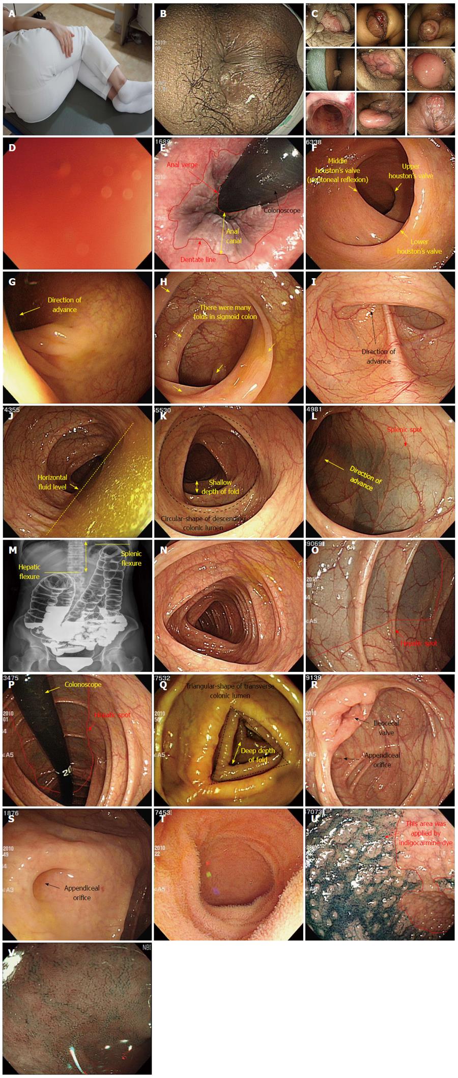
Figure 7 Procedure description.
A: For example, detailed left lateral decubitus position (the examinee with knees bent and pulled up); B: Perianal lesion; C: Various anal lesions; D: Red-out sign; E: Anal canal and distal rectum (Retroflexion view); F: rectum; G: Rectosigmoid junction; H: Sigmoid colon; I: Sigmoidodescending junction; J: Descending colon with horizontal fluid; K: Descending colon (fluid-removal view); L: Splenic flexure and spot; M: Comparison of splenic and hepatic flexures (barium study view); N: Transverse colon; O: Hepatic flexure and spot; P: Hepatic flexure and spot (retroflexion view); Q: Ascending colon; R: Cecum and ileocecal valve; S: Cecal base and appendix orifice; T, U, V: Terminal ileum (water filling view, indigocarmine-dye view, narrow-band imaging view, respectively).
- Citation: Lee SH, Park YK, Lee DJ, Kim KM. Colonoscopy procedural skills and training for new beginners. World J Gastroenterol 2014; 20(45): 16984-16995
- URL: https://www.wjgnet.com/1007-9327/full/v20/i45/16984.htm
- DOI: https://dx.doi.org/10.3748/wjg.v20.i45.16984


