Copyright
©2014 Baishideng Publishing Group Inc.
World J Gastroenterol. Dec 7, 2014; 20(45): 16881-16890
Published online Dec 7, 2014. doi: 10.3748/wjg.v20.i45.16881
Published online Dec 7, 2014. doi: 10.3748/wjg.v20.i45.16881
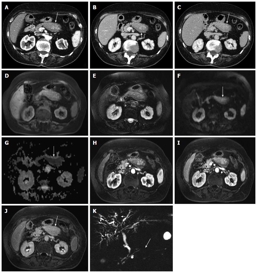
Figure 2 Focal-type autoimmune pancreatitis.
A-C: Computed tomography: the body of the pancreas appears focally enlarged (arrow in A) with a hypodense peripancreatic rim, better visible in the venous phase (arrow in B). The lesion shows fair enhancement resulting almost isodense in the delayed phase (arrow in C); D-K: Magnetic resonance: the affected portion of the pancreas is slightly hypointense on T1-weighted fat-saturated (arrow in D) images and slightly hyperintense on T2-weighted fat-saturated images (E), with diffusion coefficient restriction (arrows in F-G) with intermediate-high b values. At dynamic examination the pancreatic lesion shows fair enhancement resulting almost isodense in the delayed phase (arrow in J). At magnetic resonance cholangiopancr-eatography the main pancreatic duct shows a focal stenosis (long arrow in K) without upstream dilation. The intrahepatic bile ducts present irregular slightly stenotic portions (short arrows in K), due to involvement in the autoimmune process.
- Citation: Crosara S, D'Onofrio M, De Robertis R, Demozzi E, Canestrini S, Zamboni G, Pozzi Mucelli R. Autoimmune pancreatitis: Multimodality non-invasive imaging diagnosis. World J Gastroenterol 2014; 20(45): 16881-16890
- URL: https://www.wjgnet.com/1007-9327/full/v20/i45/16881.htm
- DOI: https://dx.doi.org/10.3748/wjg.v20.i45.16881


