Copyright
©2014 Baishideng Publishing Group Inc.
World J Gastroenterol. Dec 7, 2014; 20(45): 16858-16867
Published online Dec 7, 2014. doi: 10.3748/wjg.v20.i45.16858
Published online Dec 7, 2014. doi: 10.3748/wjg.v20.i45.16858
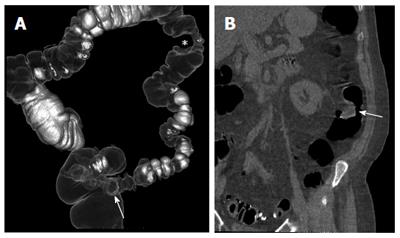
Figure 2 Patient with incomplete colonoscopy due to severe angulation and stricture secondary to diverticular disease of the sigmoid colon.
A: On a volume-rendered colon map, a stricture of the sigmoid colon (arrow) and a large filling defect (asterisk) on the medial wall of the descending colon are evident; B: On a coronal image, a large polyp (arrow) of the descending colon is observed.
- Citation: Laghi A. Computed tomography colonography in 2014: An update on technique and indications. World J Gastroenterol 2014; 20(45): 16858-16867
- URL: https://www.wjgnet.com/1007-9327/full/v20/i45/16858.htm
- DOI: https://dx.doi.org/10.3748/wjg.v20.i45.16858


