Copyright
©2014 Baishideng Publishing Group Inc.
World J Gastroenterol. Nov 28, 2014; 20(44): 16480-16488
Published online Nov 28, 2014. doi: 10.3748/wjg.v20.i44.16480
Published online Nov 28, 2014. doi: 10.3748/wjg.v20.i44.16480
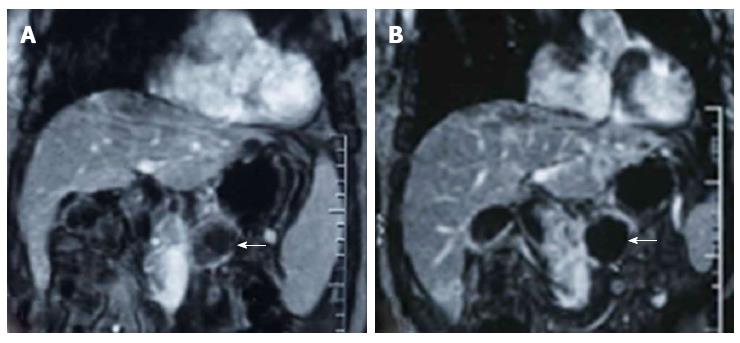
Figure 3 Contrast-enhanced T-weighted MR images obtained in a patient treated with high intensity focused ultrasound for advanced pancreatic cancer.
The tumor was 4.5 cm in diameter and located in the body of the pancreas. A: Image obtained before high intensity focused ultrasound (HIFU) shows blood supply in the pancreatic lesion (arrows); B: Image obtained 2 wk after HIFU shows no evidence of contrast enhancement in the treated lesion (arrows), which is indicative of complete coagulation necrosis in the treated pancreatic cancer.
- Citation: Wu F. High intensity focused ultrasound: A noninvasive therapy for locally advanced pancreatic cancer. World J Gastroenterol 2014; 20(44): 16480-16488
- URL: https://www.wjgnet.com/1007-9327/full/v20/i44/16480.htm
- DOI: https://dx.doi.org/10.3748/wjg.v20.i44.16480


