Copyright
©2014 Baishideng Publishing Group Inc.
World J Gastroenterol. Nov 21, 2014; 20(43): 16377-16380
Published online Nov 21, 2014. doi: 10.3748/wjg.v20.i43.16377
Published online Nov 21, 2014. doi: 10.3748/wjg.v20.i43.16377
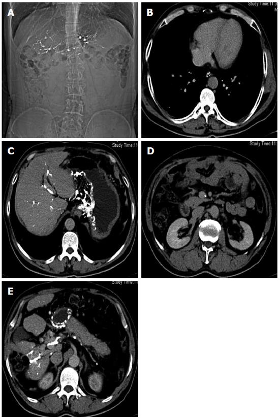
Figure 1 Plain abdominal radiography revealed linear and track-like calcification.
A: Abdominal plain film showing well-defined linear and track-like calcification, with irregular margins directed along the course of the portal vein; B: Computed tomography (CT) scan showing speckled calcifications in the esophageal vein; C: Linear calcification along the portal vein wall and globular calcification around the lesser curvature of stomach were found on CT images; D: CT scan revealed speckled calcification along the left wall of the superior mesenteric vein; E: Dense calcifications along the splenic vein were present on axial CT imaging.
- Citation: Wang CE, Sun CJ, Huang S, Wang YH, Xie LL. Extensive calcifications in portal venous system in a patient with hepatocarcinoma. World J Gastroenterol 2014; 20(43): 16377-16380
- URL: https://www.wjgnet.com/1007-9327/full/v20/i43/16377.htm
- DOI: https://dx.doi.org/10.3748/wjg.v20.i43.16377


