Copyright
©2014 Baishideng Publishing Group Inc.
World J Gastroenterol. Nov 21, 2014; 20(43): 16359-16363
Published online Nov 21, 2014. doi: 10.3748/wjg.v20.i43.16359
Published online Nov 21, 2014. doi: 10.3748/wjg.v20.i43.16359
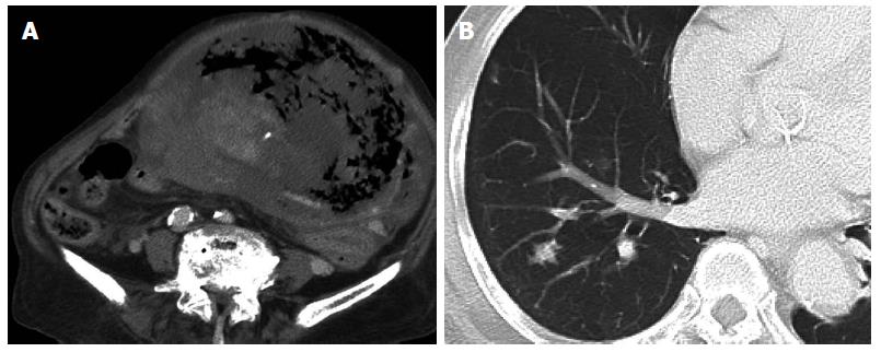
Figure 1 Preoperative computed tomography.
A: Abdominal computed tomography (CT) shows a giant tumor with central necrosis, extending from the epigastrium to the pelvic cavity; B: Chest CT shows multiple lung metastases.
- Citation: Takahashi M, Ohara M, Kimura N, Domen H, Yamabuki T, Komuro K, Tsuchikawa T, Hirano S, Iwashiro N. Giant primary angiosarcoma of the small intestine showing severe sepsis. World J Gastroenterol 2014; 20(43): 16359-16363
- URL: https://www.wjgnet.com/1007-9327/full/v20/i43/16359.htm
- DOI: https://dx.doi.org/10.3748/wjg.v20.i43.16359


