Copyright
©2014 Baishideng Publishing Group Inc.
World J Gastroenterol. Nov 21, 2014; 20(43): 16191-16196
Published online Nov 21, 2014. doi: 10.3748/wjg.v20.i43.16191
Published online Nov 21, 2014. doi: 10.3748/wjg.v20.i43.16191
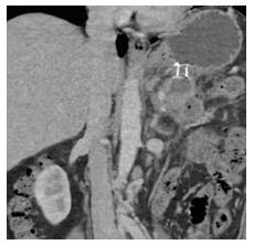
Figure 2 Computed tomography abdomen with visible metal stent between stomach and residual walled-off necrosis 6 wk after endoscopic ultrasonography guided insertion (initial cyst diameter 14 cm).
- Citation: Braden B, Dietrich CF. Endoscopic ultrasonography-guided endoscopic treatment of pancreatic pseudocysts and walled-off necrosis: New technical developments. World J Gastroenterol 2014; 20(43): 16191-16196
- URL: https://www.wjgnet.com/1007-9327/full/v20/i43/16191.htm
- DOI: https://dx.doi.org/10.3748/wjg.v20.i43.16191


