Copyright
©2014 Baishideng Publishing Group Inc.
World J Gastroenterol. Nov 14, 2014; 20(42): 15925-15930
Published online Nov 14, 2014. doi: 10.3748/wjg.v20.i42.15925
Published online Nov 14, 2014. doi: 10.3748/wjg.v20.i42.15925
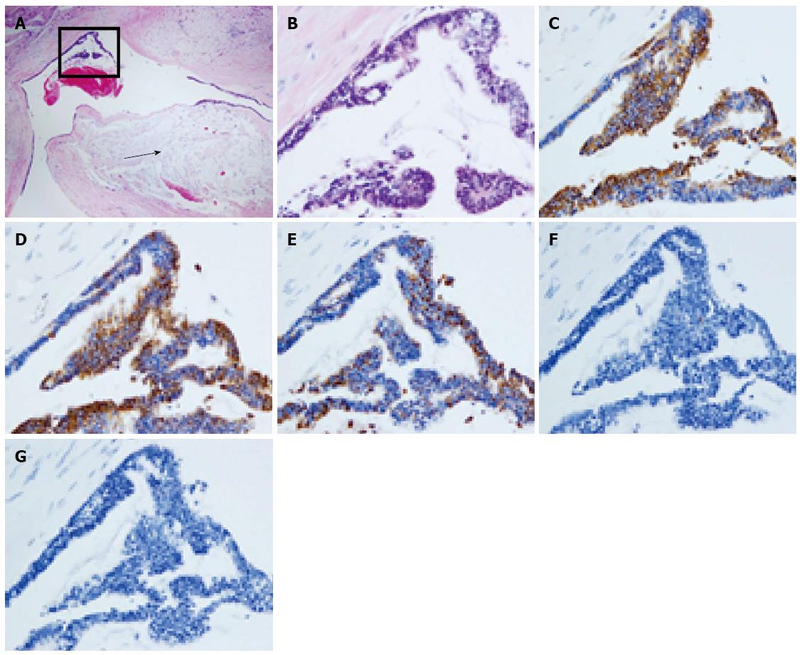
Figure 7 Tumors have proliferated and have a partly papillary appearance.
A: Secreting mucin into the parenchyma (arrow) (HE stain × 40); B: HE × 200; C: CK7 positive; D: CK20 positive; E: MUC2 positive; F: MUC5AC negative; G: MUC6 negative.
- Citation: Hachiya H, Kita J, Shiraki T, Iso Y, Shimoda M, Kubota K. Intraductal papillary neoplasm of the bile duct developing in a patient with primary sclerosing cholangitis: A case report. World J Gastroenterol 2014; 20(42): 15925-15930
- URL: https://www.wjgnet.com/1007-9327/full/v20/i42/15925.htm
- DOI: https://dx.doi.org/10.3748/wjg.v20.i42.15925


