Copyright
©2014 Baishideng Publishing Group Inc.
World J Gastroenterol. Nov 14, 2014; 20(42): 15616-15623
Published online Nov 14, 2014. doi: 10.3748/wjg.v20.i42.15616
Published online Nov 14, 2014. doi: 10.3748/wjg.v20.i42.15616
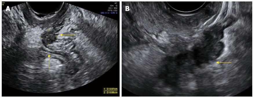
Figure 1 Transvaginal ultrasound image, sagittal view.
A: Hypoechoic nodule in the rectovaginal septum measuring 1 cm × 0.9 cm (arrow). The nodule obliterates the pouch of Douglas, invades the anterior rectal wall, and causes anatomical distorsion (dotted arrow); B: A large hypoechoic retrocervical nodule affecting the rectosigmoid colon is seen (arrow). Note the typical “indian headdress sign”, indicating deep endometriosis into the bowel wall.
- Citation: Wolthuis AM, Meuleman C, Tomassetti C, D’Hooghe T, de Buck van Overstraeten A, D’Hoore A. Bowel endometriosis: Colorectal surgeon’s perspective in a multidisciplinary surgical team. World J Gastroenterol 2014; 20(42): 15616-15623
- URL: https://www.wjgnet.com/1007-9327/full/v20/i42/15616.htm
- DOI: https://dx.doi.org/10.3748/wjg.v20.i42.15616


