Copyright
©2014 Baishideng Publishing Group Inc.
World J Gastroenterol. Nov 7, 2014; 20(41): 15462-15466
Published online Nov 7, 2014. doi: 10.3748/wjg.v20.i41.15462
Published online Nov 7, 2014. doi: 10.3748/wjg.v20.i41.15462
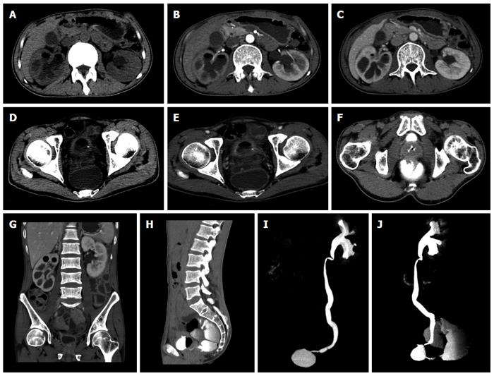
Figure 1 Computed tomography images indicated the inflammatory lesions of the right kidney, air gas in the right renal calyx and thickened wall of the right ureter (A-C, G), left ureto-cystic stone (D, E) and the vesico-rectal fistula (F, H-J).
A: Plain computed tomography (CT) scan showed enlargement of the right kidney with rounded lesions of low density in the parenchyma and air gas in the renal calyx and hydronephrosis of the left kidney; B: No enhancement of the lesions in the arterial phase; C: No enhancement of the lesions in the venous phase; D: Plain CT scan showed air gas in the bladder, thickened wall of the contracted bladder and left ureto-cystic stone; E: No enhancement of the lesions in the venous phase; F: Axial CT scan image of CT urography (CTU) showed the vesico-rectal fistula; G: Coronal 2D reconstruction image showed enlargement of the right kidney with rounded lesions of low density in the parenchyma and air gas in the renal calyx and thickened ureter wall; H: Sagittal 2D reconstruction image of CTU showed the vesico-rectal fistula, contrast medium in the rectum and air gas in the contracted bladder; I: Coronal 3D reconstruction image of CTU showed left hydronephrosis, contracted bladder and loss of function of the right kidney; J: Sagittal 3D reconstruction image of CTU showed the vesico-rectal fistula, contrast medium in the rectum, the left hydronephrosis, contracted bladder and loss of function of the right kidney.
- Citation: Wei XQ, Zou Y, Wu ZE, Abassa KK, Mao W, Tao J, Kang Z, Wen ZF, Wu B. Acute diarrhea and metabolic acidosis caused by tuberculous vesico-rectal fistula. World J Gastroenterol 2014; 20(41): 15462-15466
- URL: https://www.wjgnet.com/1007-9327/full/v20/i41/15462.htm
- DOI: https://dx.doi.org/10.3748/wjg.v20.i41.15462


