Copyright
©2014 Baishideng Publishing Group Inc.
World J Gastroenterol. Nov 7, 2014; 20(41): 15454-15461
Published online Nov 7, 2014. doi: 10.3748/wjg.v20.i41.15454
Published online Nov 7, 2014. doi: 10.3748/wjg.v20.i41.15454
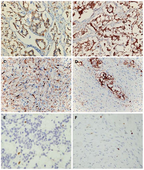
Figure 5 Immunohistochemical analysis of the lesions in the duodenum, lymph nodes and pelvic cavity.
The epithelioid cells were positive for cytokeratin (AE1/AE3) (A) and chromogranin A (B). The spindle-shaped cells were diffusely positive for S-100 protein (C), and ganglion-like cells were positive for synaptophysin (D). Ki-67 index was low in either epithelioid cell areas (E) or spindle-shaped cell areas (F). A-D: Immunohistochemical staining with original magnification × 400.
- Citation: Li B, Li Y, Tian XY, Luo BN, Li Z. Malignant gangliocytic paraganglioma of the duodenum with distant metastases and a lethal course. World J Gastroenterol 2014; 20(41): 15454-15461
- URL: https://www.wjgnet.com/1007-9327/full/v20/i41/15454.htm
- DOI: https://dx.doi.org/10.3748/wjg.v20.i41.15454


