Copyright
©2014 Baishideng Publishing Group Inc.
World J Gastroenterol. Nov 7, 2014; 20(41): 15454-15461
Published online Nov 7, 2014. doi: 10.3748/wjg.v20.i41.15454
Published online Nov 7, 2014. doi: 10.3748/wjg.v20.i41.15454
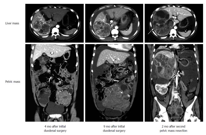
Figure 2 Postoperative radiological findings in follow-up periods.
Computed tomography (CT) scans at 4 mo and 9 mo after initial duodenal surgery showed distant metastatic lesions located in the liver (white arrows) and pelvic cavity (black arrows). The mass in the pelvic cavity became larger and pressed the surrounding tissues. Two months after second pelvic surgery, CT scans showed that the mass re-grew in the pelvic cavity (black arrows) and the liver mass became larger (white arrows).
- Citation: Li B, Li Y, Tian XY, Luo BN, Li Z. Malignant gangliocytic paraganglioma of the duodenum with distant metastases and a lethal course. World J Gastroenterol 2014; 20(41): 15454-15461
- URL: https://www.wjgnet.com/1007-9327/full/v20/i41/15454.htm
- DOI: https://dx.doi.org/10.3748/wjg.v20.i41.15454


