Copyright
©2014 Baishideng Publishing Group Inc.
World J Gastroenterol. Nov 7, 2014; 20(41): 15367-15373
Published online Nov 7, 2014. doi: 10.3748/wjg.v20.i41.15367
Published online Nov 7, 2014. doi: 10.3748/wjg.v20.i41.15367
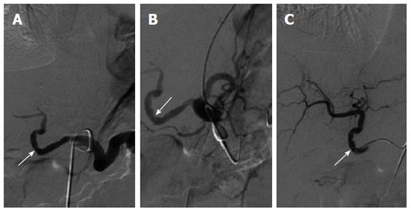
Figure 2 Radiological images during occluding the splenic artery (arrow is the anastomotic site).
A: Selective celiac trunk arteriogram shows reduced hepatic artery perfusion and enlarged splenic artery perfusion; B: Increased hepatic arterial perfusion was seen after occluding the splenic artery temporarily; C: Normal hepatic arterial flow was reestablished 5 d after the treatment.
- Citation: Song JY, Shi BY, Zhu ZD, Zheng DH, Li G, Feng LK, Zhou L, Wu TT, Du GS. New strategies for prevention and treatment of splenic artery steal syndrome after liver transplantation. World J Gastroenterol 2014; 20(41): 15367-15373
- URL: https://www.wjgnet.com/1007-9327/full/v20/i41/15367.htm
- DOI: https://dx.doi.org/10.3748/wjg.v20.i41.15367


