Copyright
©2014 Baishideng Publishing Group Inc.
World J Gastroenterol. Nov 7, 2014; 20(41): 15216-15232
Published online Nov 7, 2014. doi: 10.3748/wjg.v20.i41.15216
Published online Nov 7, 2014. doi: 10.3748/wjg.v20.i41.15216
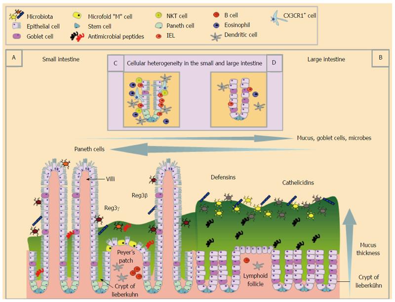
Figure 1 Schematic representation of the structural and cellular heterogeneity within the small and large intestine during homeostatic conditions.
The small (A and C) and large (B and D) intestine differ greatly in their structure and cellular composition. The small intestinal epithelium is folded to create crypts of Lieberkühn, has finger-like projections called villi and epithelial cell possess striated microvilli (A) whereas the large intestine (B) has crypts and lacks villi. The small intestine has Peyer’s patches, the main site of Microfold or “M” cells that transcytose luminal particulate antigens that are rare in the larger intestine. Both the small and large intestine are covered by mucus, which is much thicker in the large intestine (B) than in the small intestine (A). The large intestine houses the largest and most diverse microbial populations of the two sites (B). Anti-microbial peptides are also differentially expressed along the gastrointestinal tract. The distribution of immune cells also differ in the small and large intestine (inset C and D) with Natural killer T cells, Intraepithelial lymphocytes (IEL), eosinophils and dendritic cells found in the highest numbers in the small intestinal epithelium under steady state conditions. Cell populations are defined in the key.
- Citation: Bowcutt R, Forman R, Glymenaki M, Carding SR, Else KJ, Cruickshank SM. Heterogeneity across the murine small and large intestine. World J Gastroenterol 2014; 20(41): 15216-15232
- URL: https://www.wjgnet.com/1007-9327/full/v20/i41/15216.htm
- DOI: https://dx.doi.org/10.3748/wjg.v20.i41.15216


