Copyright
©2014 Baishideng Publishing Group Inc.
World J Gastroenterol. Oct 28, 2014; 20(40): 14992-14996
Published online Oct 28, 2014. doi: 10.3748/wjg.v20.i40.14992
Published online Oct 28, 2014. doi: 10.3748/wjg.v20.i40.14992
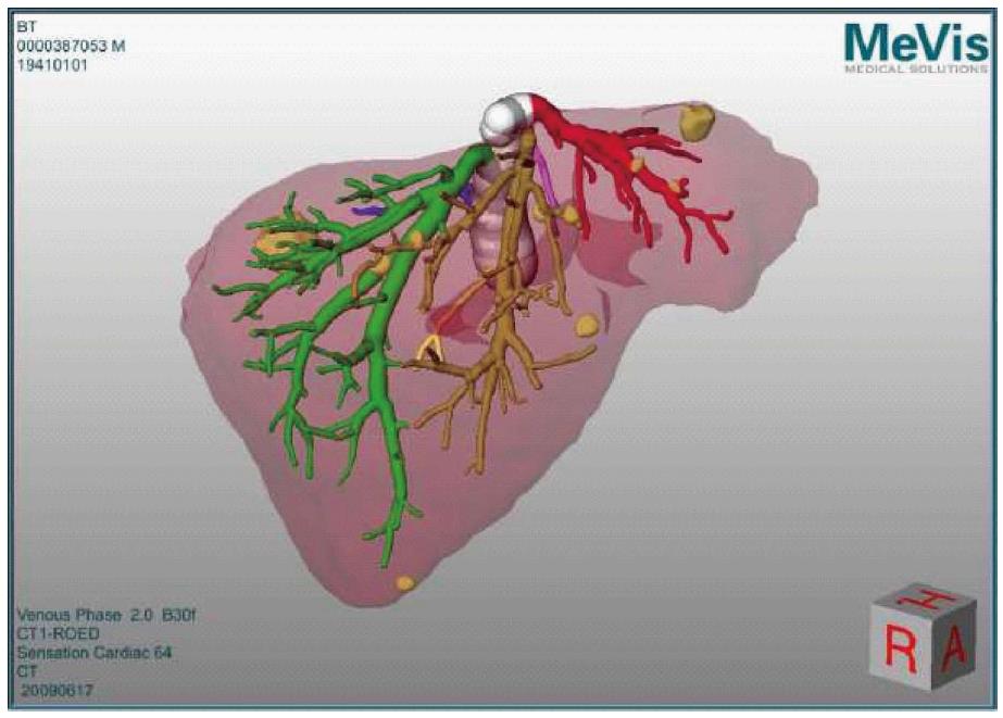
Figure 1 Planning data (as obtained from MeVis) depicting the 3D liver model as calculated from the preoperative triphasic computed tomography scan.
The image shows the hepatic veins (left hepatic vein in red, middle hepatic vein in orange-brown, right hepatic vein in green). The tumors are seen as yellow masses, distributed in both liver lobes.
- Citation: Banz VM, Baechtold M, Weber S, Peterhans M, Inderbitzin D, Candinas D. Computer planned, image-guided combined resection and ablation for bilobar colorectal liver metastases. World J Gastroenterol 2014; 20(40): 14992-14996
- URL: https://www.wjgnet.com/1007-9327/full/v20/i40/14992.htm
- DOI: https://dx.doi.org/10.3748/wjg.v20.i40.14992


