Copyright
©2014 Baishideng Publishing Group Inc.
World J Gastroenterol. Oct 28, 2014; 20(40): 14831-14840
Published online Oct 28, 2014. doi: 10.3748/wjg.v20.i40.14831
Published online Oct 28, 2014. doi: 10.3748/wjg.v20.i40.14831
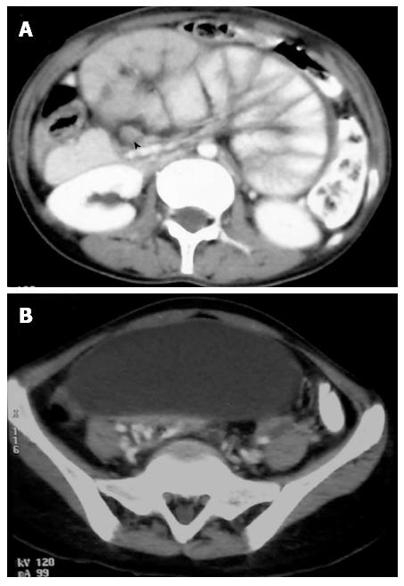
Figure 5 Computed tomography images.
A: Axial images of the same patient showing small bowel loops congregated in the centre of the abdomen encased by a soft tissue density membrane. Enlarged discrete lymph nodes are also seen in the mesentery (arrow head); B: Loculated ascites is seen just below the level of conglomerate bowel loops.
- Citation: Debi U, Ravisankar V, Prasad KK, Sinha SK, Sharma AK. Abdominal tuberculosis of the gastrointestinal tract: Revisited. World J Gastroenterol 2014; 20(40): 14831-14840
- URL: https://www.wjgnet.com/1007-9327/full/v20/i40/14831.htm
- DOI: https://dx.doi.org/10.3748/wjg.v20.i40.14831


