Copyright
©2014 Baishideng Publishing Group Inc.
World J Gastroenterol. Oct 28, 2014; 20(40): 14760-14777
Published online Oct 28, 2014. doi: 10.3748/wjg.v20.i40.14760
Published online Oct 28, 2014. doi: 10.3748/wjg.v20.i40.14760
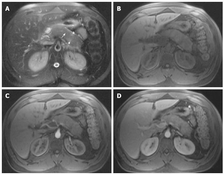
Figure 20 Autoimmune pancreatitis.
A: Axial single-shot turbo spin-echo T2-weighted (HASTE) image with fat-suppression; B: Axial T1-weighted image. Post-Gadolinium 3D-GRE T1-weighted images with fat-suppression during the hepatic arterial-dominant (C) and hepatic-venous phases (D). There is evidence of diffuse pancreatic swelling with patchy mildly increased T2 signal (arrowheads) (A), decreased T1 signal (B), loss of the normal pancreatic lobulations, and diffuse obliteration of the pancreatic duct (arrow) (A); associated with patchy decreased early enhancement (C) and progression of enhancement on subsequent phases (D) in keeping with autoimmune pancreatitis.
- Citation: Manikkavasakar S, AlObaidy M, Busireddy KK, Ramalho M, Nilmini V, Alagiyawanna M, Semelka RC. Magnetic resonance imaging of pancreatitis: An update. World J Gastroenterol 2014; 20(40): 14760-14777
- URL: https://www.wjgnet.com/1007-9327/full/v20/i40/14760.htm
- DOI: https://dx.doi.org/10.3748/wjg.v20.i40.14760


