Copyright
©2014 Baishideng Publishing Group Inc.
World J Gastroenterol. Oct 28, 2014; 20(40): 14760-14777
Published online Oct 28, 2014. doi: 10.3748/wjg.v20.i40.14760
Published online Oct 28, 2014. doi: 10.3748/wjg.v20.i40.14760
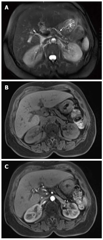
Figure 18 Chronic pancreatitis.
A: Axial single-shot turbo spin-echo T2-weighted (HASTE) image. Axial pre- (B) and post- (C) Gadolinium 3D-GRE T1-weighted image with fat-suppression during the hepatic arterial-dominant phase. There is evidence of diffuse atrophy of the pancreatic parenchyma with mild uniform pancreatic ductal dilatation (arrow) and pancreatic side-branches prominence (A), associated with diminished T1 signal intensity (B) and minimal arterial enhancement (C) in keeping with chronic pancreatitis. There are also few prominent lymph nodes in the peripancreatic and porta hepatis lymph nodes (asterisks) (A-C).
- Citation: Manikkavasakar S, AlObaidy M, Busireddy KK, Ramalho M, Nilmini V, Alagiyawanna M, Semelka RC. Magnetic resonance imaging of pancreatitis: An update. World J Gastroenterol 2014; 20(40): 14760-14777
- URL: https://www.wjgnet.com/1007-9327/full/v20/i40/14760.htm
- DOI: https://dx.doi.org/10.3748/wjg.v20.i40.14760


