Copyright
©2014 Baishideng Publishing Group Inc.
World J Gastroenterol. Oct 28, 2014; 20(40): 14760-14777
Published online Oct 28, 2014. doi: 10.3748/wjg.v20.i40.14760
Published online Oct 28, 2014. doi: 10.3748/wjg.v20.i40.14760
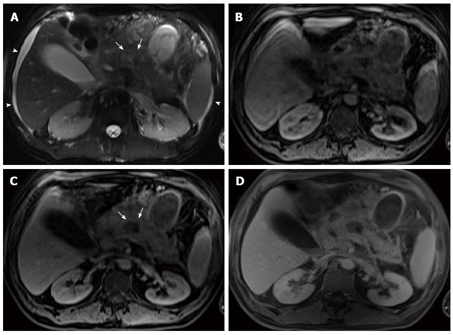
Figure 13 Infected peripancreatic fluid and interrupted duct syndrome in a patient with acute pancreatitis.
A: Axial single-shot turbo spin-echo T2-weighted (HASTE) image with fat-suppression. Axial pre- (B) and post- (C) Gadolinium 3D-GRE T1-weighted images with fat-suppression during the hepatic arterial-dominant phase; D: Axial post-Gadolinium radial 3D-GRE T1-weighted images with fat-suppression during the interstitial phase. There is a large irregular multiloculated (partially visualized) peripancreatic collection. It demonstrates decreased T2 (A) and T1 (B) signal with a thick rim of increased T2 signal (arrows) (A) that shows enhancement on delayed images (arrows, C, D) in keeping with infected necrotic collection (proven by fine needle aspiration). There is also a well-defined fluid collection in the lesser sac (asterisk) (A) with an enhancing thin wall (D) and minimal ascites (arrowheads, A). Of note is the motion robustness of the radial 3D-GRE (D) compared to convention 3D-GRE (C) sequence, facilitating imaging of sick uncooperative patients.
- Citation: Manikkavasakar S, AlObaidy M, Busireddy KK, Ramalho M, Nilmini V, Alagiyawanna M, Semelka RC. Magnetic resonance imaging of pancreatitis: An update. World J Gastroenterol 2014; 20(40): 14760-14777
- URL: https://www.wjgnet.com/1007-9327/full/v20/i40/14760.htm
- DOI: https://dx.doi.org/10.3748/wjg.v20.i40.14760


