Copyright
©2014 Baishideng Publishing Group Inc.
World J Gastroenterol. Oct 28, 2014; 20(40): 14760-14777
Published online Oct 28, 2014. doi: 10.3748/wjg.v20.i40.14760
Published online Oct 28, 2014. doi: 10.3748/wjg.v20.i40.14760
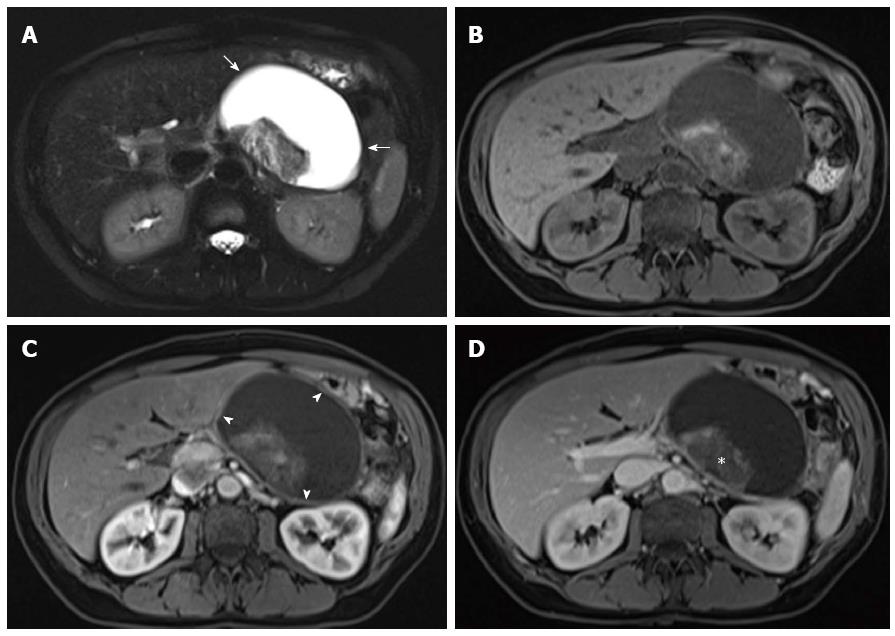
Figure 12 Necrotizing pancreatitis with peripancreatic walled-off necrosis.
A: Axial single-shot turbo spin-echo T2-weighted (HASTE) image. Axial (B) pre- and post-Gadolinium 3D-GRE T1-weighted images with fat-suppression during the hepatic arterial-dominant (C) and hepatic-venous phases (D). There is a well-defined fluid collection (arrows) at the region of the pancreatic body/tail (A, B); which demonstrates a uniform mildly enhancing thickened rim (arrowheads) (C,D) and contains a dependent non-enhancing debris (asterisk) (C, D) in keeping with necrotizing pancreatitis and walled-off necrosis.
- Citation: Manikkavasakar S, AlObaidy M, Busireddy KK, Ramalho M, Nilmini V, Alagiyawanna M, Semelka RC. Magnetic resonance imaging of pancreatitis: An update. World J Gastroenterol 2014; 20(40): 14760-14777
- URL: https://www.wjgnet.com/1007-9327/full/v20/i40/14760.htm
- DOI: https://dx.doi.org/10.3748/wjg.v20.i40.14760


